PET/CT examination
Comprehensive diagnostics through full body scan
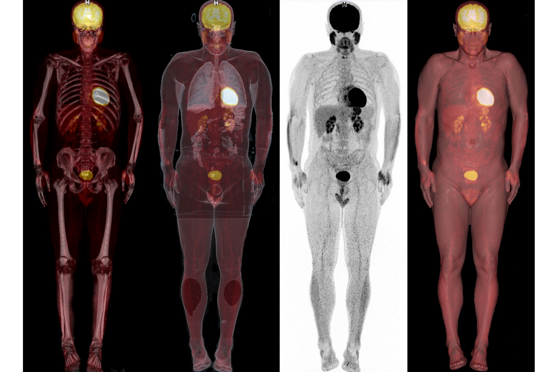
A PET/CT examinamion combines the strengths of functional imaging (positron emission tomography (PET)) with the anatomical cross-sectional images of a computer tomography (CT) so that changes in your body can be both detected and precisely localized.
PET/CT is a whole-body examination. This is particularly relevant in oncology, as tumors in suspected areas and distant metastases can be examined and imaged with the aid of a single examination. PET/CT is characterized in particular by the fact that even the smallest tumors and metastases can be clearly imaged.
In addition to oncological diagnostics, PET/CT is regularly used for questionable neurological cases in order to detect anatomical and functional changes in the brain. PET/CT has proven particularly useful in diagnosing Alzheimer’s and Parkinsons diseases.

Duration of Examination
Approx. 4 hours

Areas of application
Oncology & Neurology

Health insurance benefit
Partial
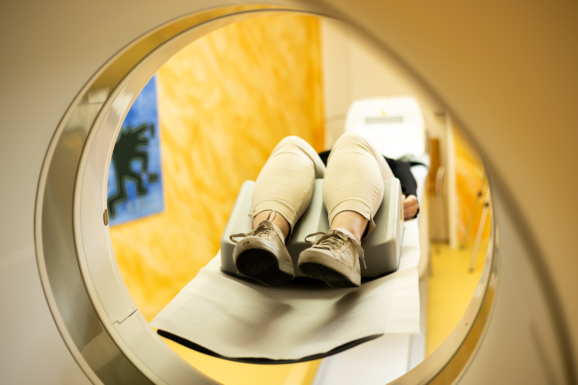
Examination procedure
Although a PET/CT examination takes an average of 25 minutes, you should allow about 4 hours for your stay with us. In the following pages, we would like to give you an overview of the examination procedure. In the case of prostate carcinoma and in other individual cases, however, there may be deviations in the procedure.


PET/CT is used in all phases of cancer diagnostics (oncology):
- Early detection
- Search for the primary cancer site
- Characterization and staging of the disease
- Therapy control of chemotherapy or radiotherapy
- Follow-up care after cancer therapy to detect relapses (recurrence diagnostics)
PET/CT is used especially for neurological diseases. Among other things, special tracers (e.g., Florbetaben) are used to detect plaques in the brain characteristic to Alzheimer’s disease with dementia.
PET/CT examinations also have high diagnostic value in answering other neurological questions such as the differentiation between depression and dementia or in degenerative multisystem diseases.
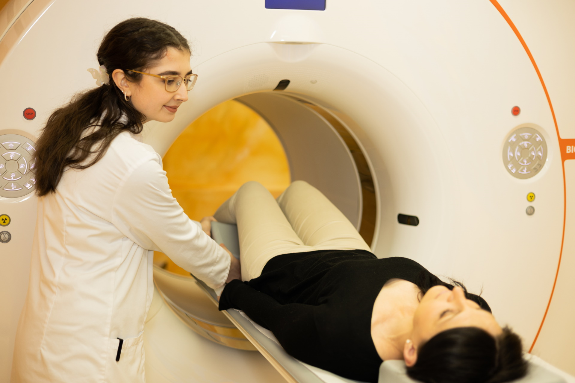
You must have an empty stomach in order for the PET/CT exam to be performed correctly.
To ensure a clear image, you must remove jewelry and other metal objects, such as clothing containing metal (e.g., buttons) or belts, before the examination. If necessary, please make sure that clothing and shoes can be easily taken off and put back on.
For privately insured patients, the examination billed according to the Gebührenordnung für Ärzte (GOÄ) is usually reimbursed.
Statutory health insurance companies, on the other hand, only cover the costs for selected indications.
In addition, there is still the possibility to privately finance a PET/CT examination. Please contact us for further information.
PET/CT at a glance
The first PET/CT arrived in our country in 2003 as the first device in all of Europe. Although the method provides an important tool for oncology and neurology, even today, only a few medical institutions offer PET/CT examinations.
The weak radioactively labeled tracer injected for the examination increasingly metabolizes and accumulates accordingly in tumors and areas of inflammation. PET can then visually display the increased glucose metabolism.
Meanwhile, computed tomography is a special X-ray procedure that uses computers to convert different absorptions of X-rays in the body into anatomically correct cross-sectional images. Fusing the PET and CT images provides precise anatomical assignment of activities detected by the PET examination.
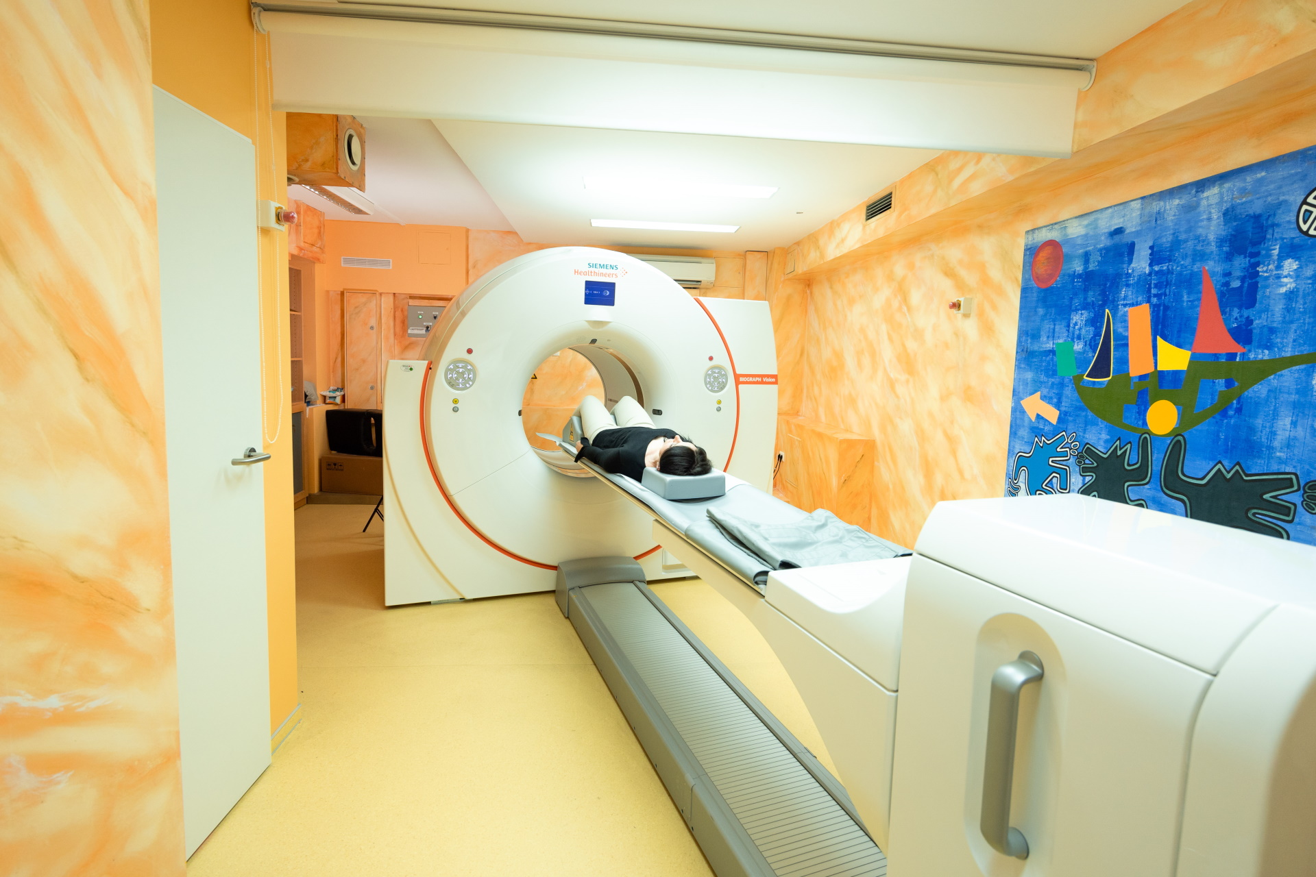
Our device
Since January 2021, we have been working with the Siemens Biograph Vision 450, a second-generation digital PET/CT with a 128-Slice CT (Edge), that uses innovative technology and a new design of LSO crystal detectors to enable higher-resolution and more precise imaging, while shortening the examination time and reducing patient radiation exposure. Wider than average with a diameter of 78cm and typically short at a length of only 136cm, the gantry (tube) facilitates a flexible and comfortable patient position. Together with the short examination times, these measurements ensure exceptional patient comfort.
Arrival of the Biograph Vision 450 at MCB
VIDEO: https://www.youtube.com/watch?v=pZMivTQCVZQ