Ultrasound examination
Sonographies – risk-free diagnostics thanks to proven method
Ultrasound waves have been used in medicine since the 1940s to examine organs and tissues. Mainly known from its use during pregnancy, ultrasound (sonography) can be applied to other parts of the body to gather information on the size and shape of the organs as well as tissue composition and changes.
Sonographies are completely risk-free, as neither radioactive tracers nor contrast media are needed. In addition, the procedure is very short, which means that you will get results quickly. Our doctors have extensive experience and are specially trained to detect even small tissue changes.
Sonograms are used in thyroid examinations and can be implemented to check for gallstones, hernias or lipomas. You can find more information about your examination options here.

Duration of examination
10 – 30 minutes

Areas of application
Sonography thyroid, abdomen, chest, neck, joints and pelvis

Health insurance benefit
Yes
Since the transducer of the ultrasound scanner lies directly on the skin, make sure to wear clothing that makes it easy to expose the corresponding part of the body for the examination (e.g., do not wear a turtleneck sweater). Further information on the examination procedure and how your can prepare will be provided by our specialist staff in advance of your sonography.
Sonographies are usually covered or reimbursed by public and private health insurance.
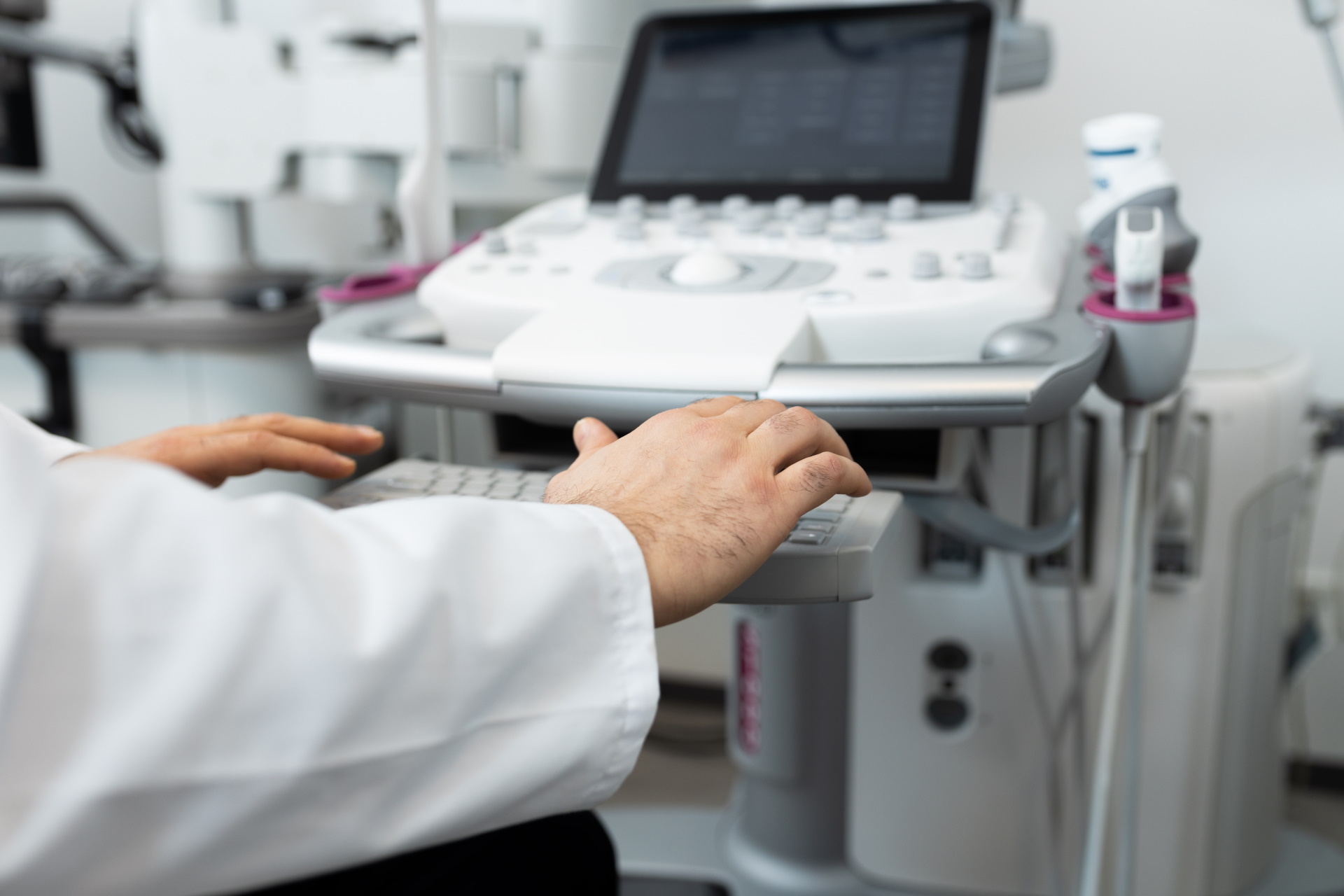
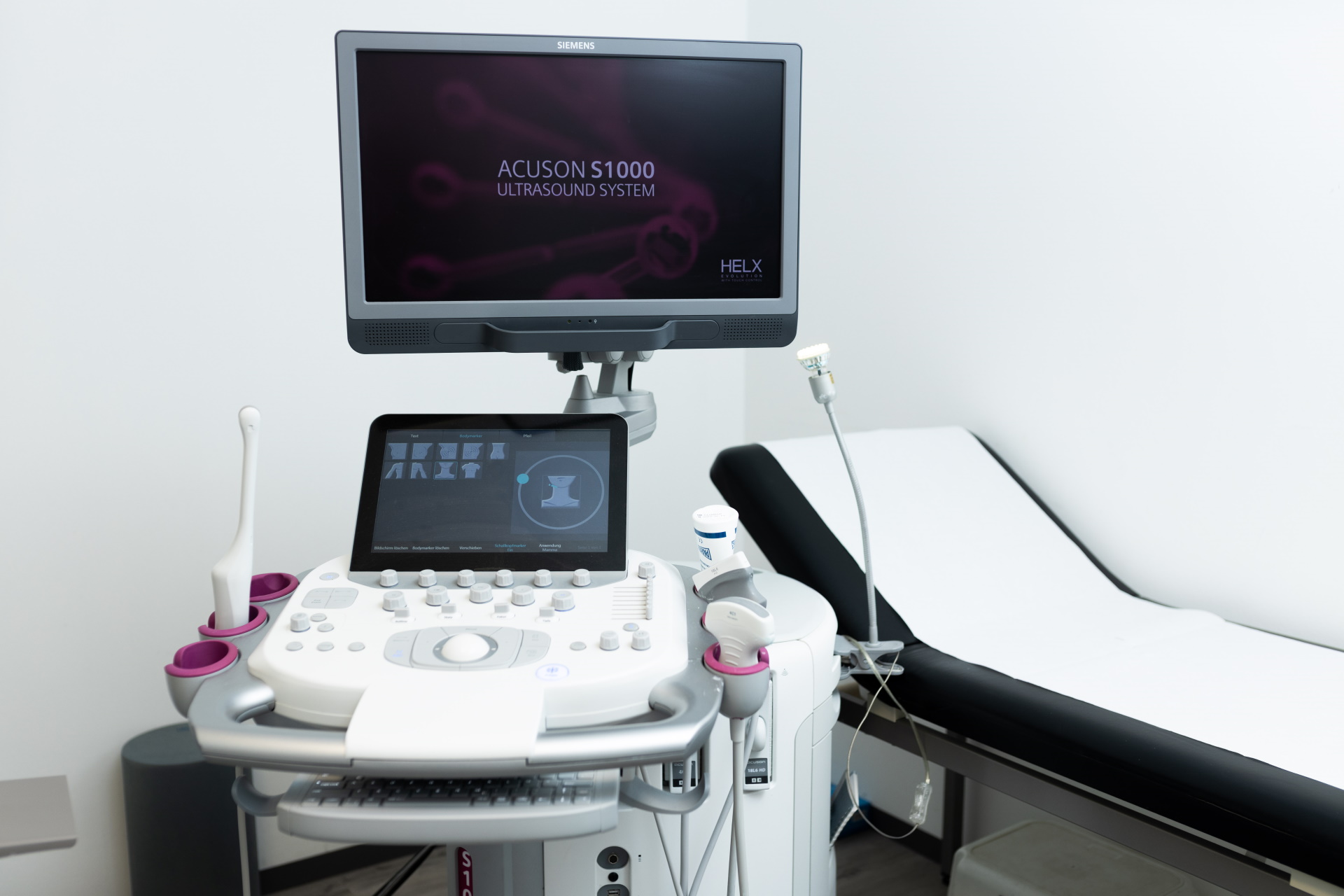
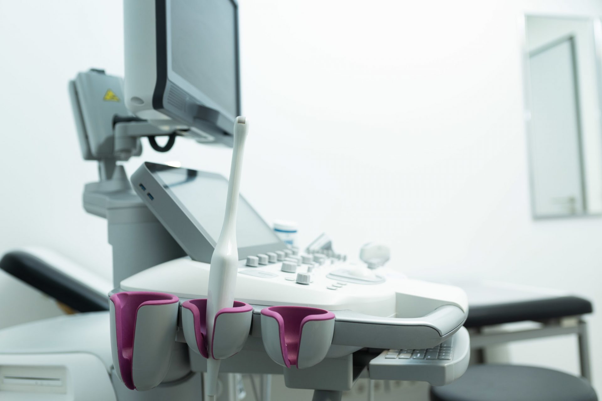
An ultrasound device works with sound waves that emanate from a transducer and are absorbed or reflected by the body depending on the type of tissue. The reflected sound waves return to the transducer, which also serves as a receiver. It converts the incoming sound waves into electrical impulses, amplifies them and transmits the information to a connected monitor. The resulting two-dimensional, black-and-white images show the size, shape and structure of the region being examined so our doctors can detect any pathological changes in the organ or tissue.