CT scan
Detailed diagnostics thanks to state-of-the-art technology
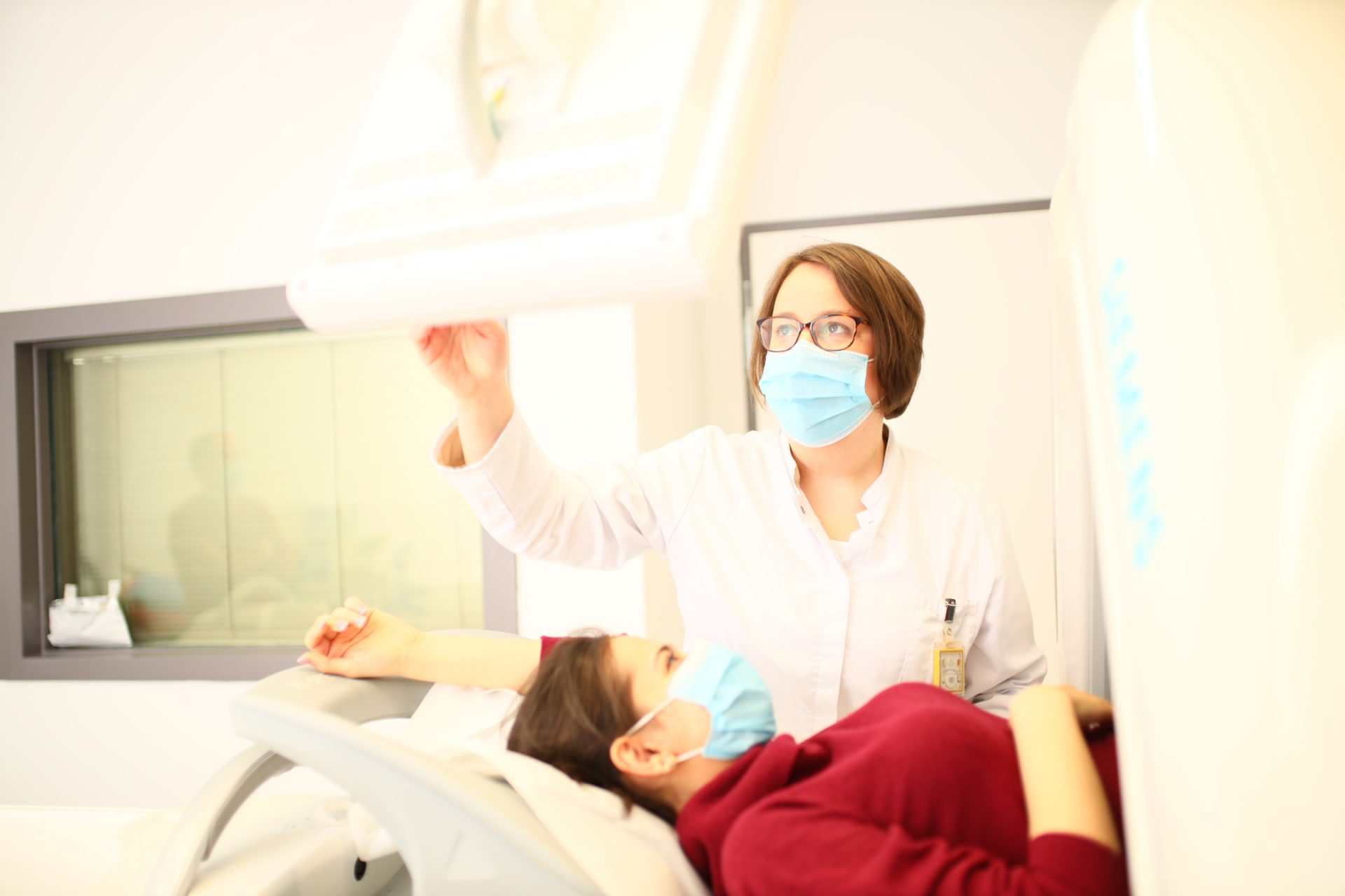
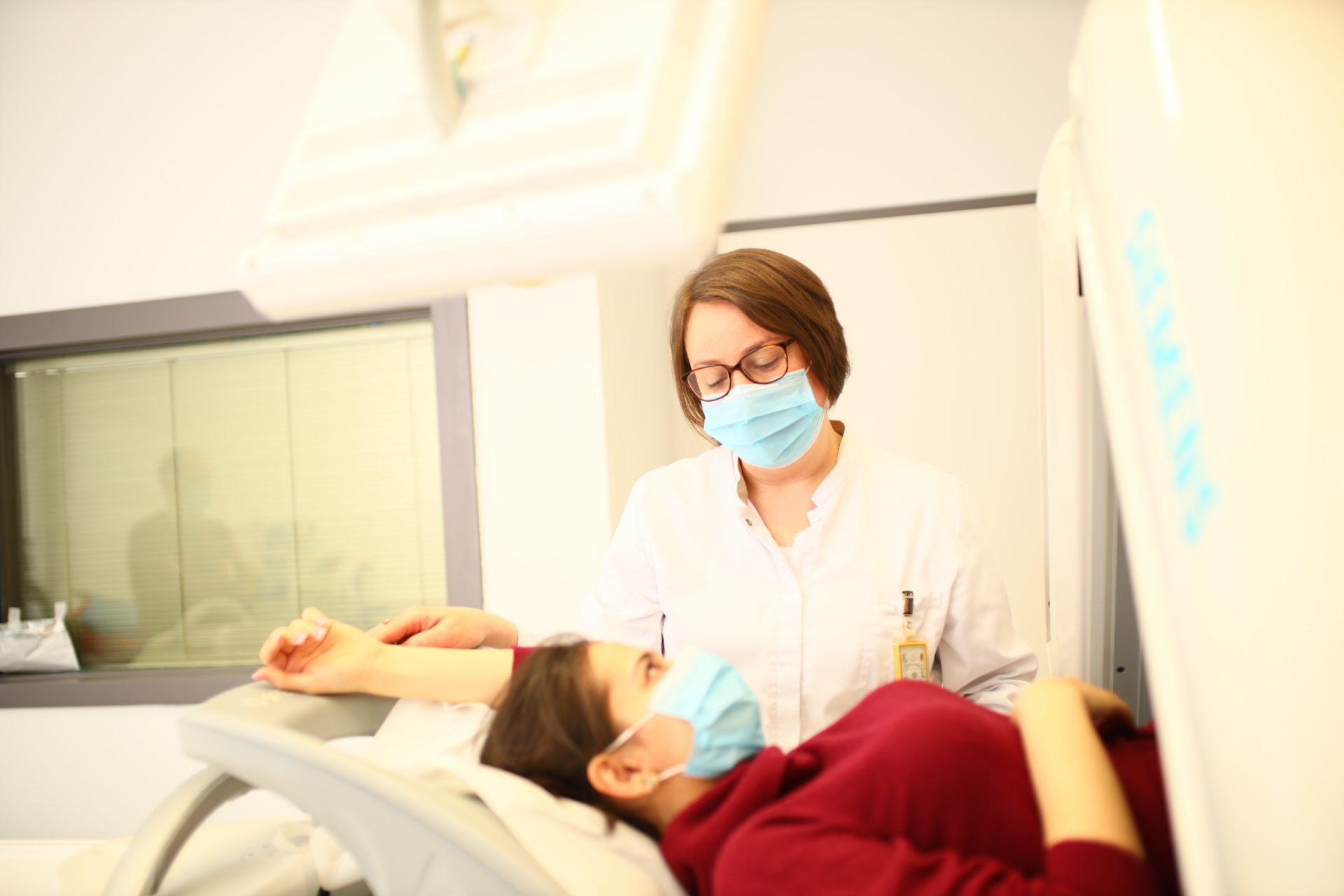
A computer tomography (CT) illustrates your body in detailed cross-sectional images so our doctors can detect even very small changes by means of a single examination. Exceptionally precise CT imagery allows for early diagnosis and a fast start to therapy. Rapid treatment increases the likelihood of a positive outcome and reduces the risk of secondary damage, as treatment can be applied in a targeted manner.
The level of detail and flexibility of the method allows for broad application.
CT technology can be used to search and locate tumors, examine the abdomen and verify lung diseases, all of which are of particular diagnostic importance. Furthermore, CT examinations are used to clarify injury or disease of bones and the skeletal system, as well as to help doctors answer neurological and cardiological questions.
For more information on the CT scans we offer, click here.
Duration of examination
Few minutes
Areas of application
Orthopedics, Oncology, Neurology & Pulmonology
Health insurence benefit
Yes, with bank transfer
Our specialist staff will provide you with all the necessary information on examination preparation and procedure in advance of your examination.
Note that all jewelry and other metal objects in the examination region must be removed before the examination in order to obtain a proper image. This includes, for example, metal-containing clothing with zippers and buttons or belts. In addition, please make sure that clothing and shoes can be taken off and put back on easily.
A CT scan is usually covered by all health insurance companies after an appropriate referral from your physician.
A computer tomograph (CT) consists of an X-ray tube and an opposing detector integrated in a ring that continuously circles you about two times per second. This movement is completely silent and imperceptible. As they pass through your body, the X-rays are attenuated or absorbed according to varying densities of tissue and bone. The rays subsequently received by the detector are converted into electronic signals from which the three-dimensional sectional images are calculated on a computer basis.


