DPX examination
Detect osteoporosis thanks to bone density measurement
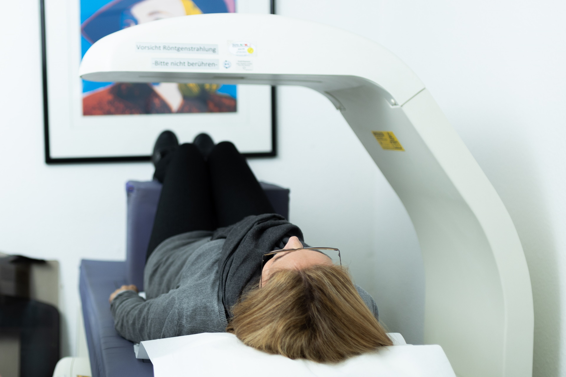
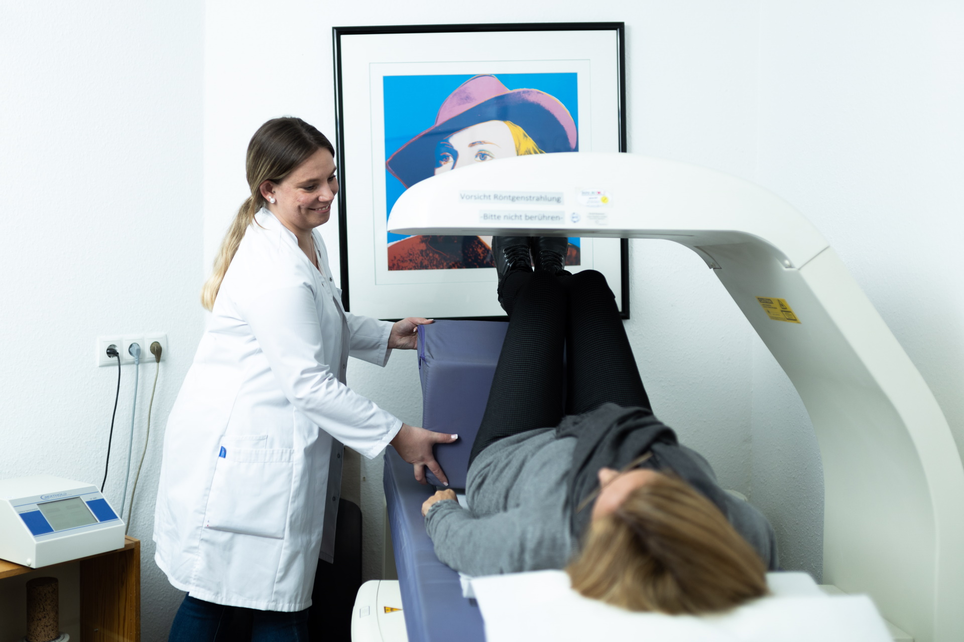
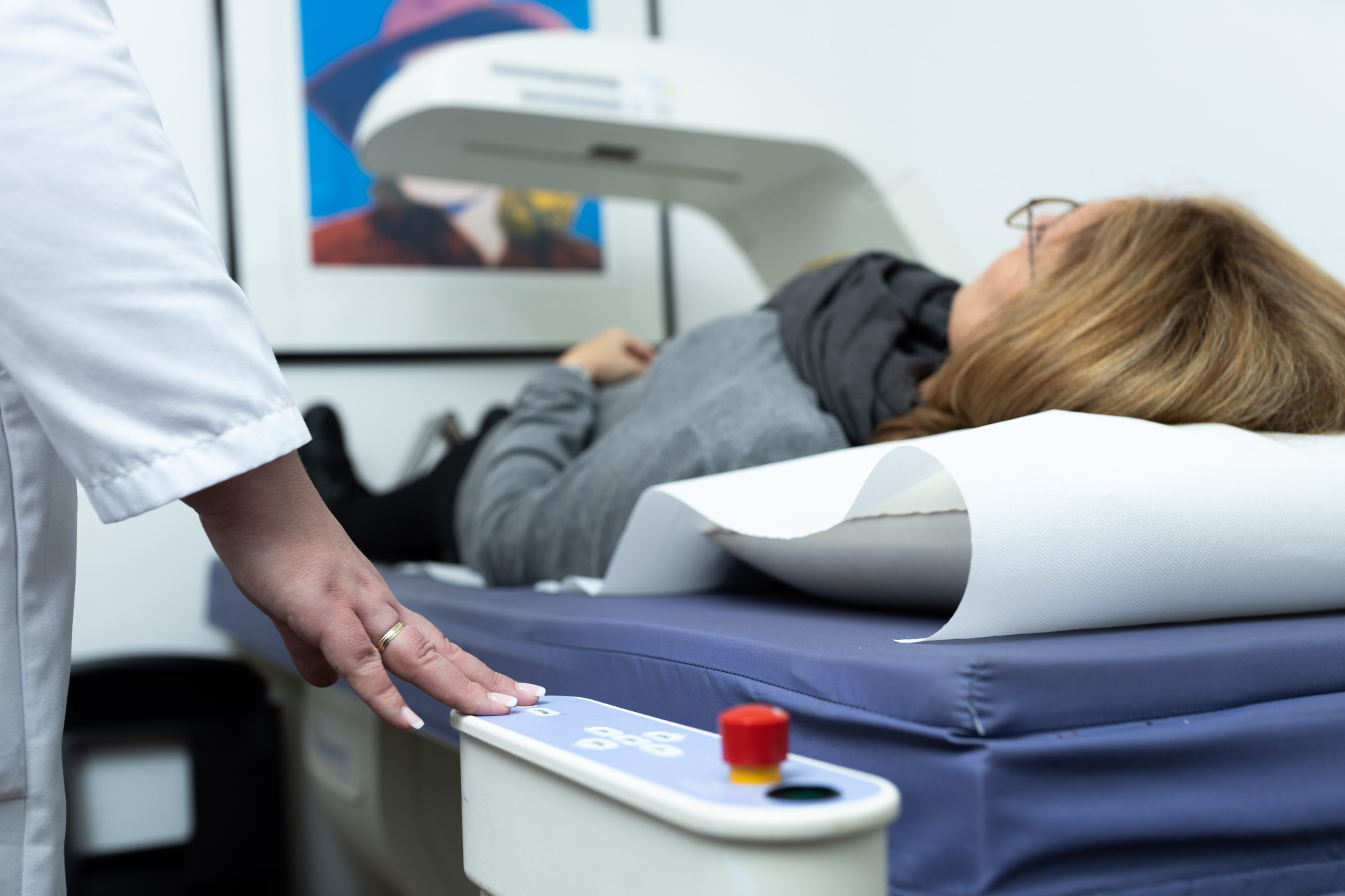
A dual photon X-ray (DPX) is used when there is an increased risk of bone density loss (osteoporosis), or when the disease has already been diagnosed.
Bone density measurement is especially important for postmenopausal women, individuals with increased alcohol or nicotine consumption or people with a family history of osteoporosis. Bone breaks (fractures) without a corresponding or related accident can also indicate osteoporosis.
A non-invasive examination, DPX uses weak X-rays which can also provide information about the condition of the bones at an early stage of the disease. Early diagnosis and regular checks during therapy make it possible to reduce the loss of bone density and thus prevent secondary damage such as frequent fractures.
Bone densitometry using DPX is the gold standard for osteoporosis diagnosis and is recommended by the German Society of Osteology.
You can also find a comprehensive overview of all our examinations here.

Duration of examination
10 minutes

Areas of application
Osteoporosis diagnostics

Health insurance benefit
Partial
Osteoporosis occurs frequently, but not exclusively, in older patients. The disease is often detected late, as many years can pass without symptoms. In this case, the earliest possible therapy is very important. You should consider a bone densitometry if one or more of the following points apply to you:
- Bone fractures without a corresponding accident
- Classic osteoporosis symptoms, such as bone pain, hunchback or clustered fractures before menopause in women
- Increased risk, e.g. heavy alcohol or nicotine consumption, family history, diabetes or malnutrition
- Post-menopausal women or those with estrogen deficiency
- Primary hyperparathyroidism (regulatory disorder of the parathyroid gland)
- Suspected osteomalacia (deficient mineral incorporation into the bones)
You do not need to make any special preparations for your DPX bone densitometry. Our specialist staff will tell you everything you need to know about the procedure in advance of your examination.
For privately insured patients, the examination billed according to the Gebührenordnung für Ärzte (GOÄ) is usually reimbursed.
The statutory health insurance funds, on the other hand, only cover the costs if there is a corresponding indication.
In addition, there is the possibility to have a DPX examination performed as a self-payer. Please contact us for further information.
A DPX examination uses X-rays generated by the X-ray tube and passed through your body. The X-rays are attenuated according to bone density. Detectors in the DPX device record the incoming radiation and its strength and forward the signal information to a connected computer. The resulting values can now be compared with other values and provide information on whether and to what extent a decrease in bone density has occurred.
How do DPX, DXA and DEXA differ?
This is always the same method. Sometimes it is also called QDR or DER.